PET (Positron Emission computed Tomography) Chinese name is "positron emission computed tomography, its clinical application history has been more than 40 years. In 1974, the first commercial PET entered the clinical, the first whole body PET in 1992 Beginning to use, with the commercialization of the whole body PET-CT with 16-row CT in 2003, the world's major manufacturers stopped the production and sales of individual PET.
As a new examination method in the nuclear medicine department, PET/CT is another revolution in the medical imaging field. Just like the Pet (pet) contained in its name, it has become the new favorite of modern nuclear medicine, so far the major hospitals in China. The Department of Nuclear Medicine has installed more than 200 units, and with the gradual reduction of market access thresholds, it is possible to enter medical insurance or part of medical insurance and domestic companies such as Lian Ying in the future to make localization efforts in large-scale medical equipment. Gradually showing the blowout situation.
While PET/CT brings surprises and gospel to the public health cause, it has caused widespread doubts and panic among professionals and the public because of its extensive and unregulated medical examinations in the past 10 years, and even some organizations exaggerating their utility propaganda. Even open or half-strong debates are intermittent. For example, the academic doctors questioned, panic, and even open or half of the heated debate. For example, the academic doctors questioned, panic, and even open or half of the heated debate. For example, the academic and medical circles have been arguing about their early ability to detect tumors and the magnitude of radiation hazards. Nowadays, the public's radiation and hazards caused by this high-force image inspection have even reached the point of "radiation".
In order to objectively and effectively make the PET-CT good equipment better for the appropriate patients, this article will open the mystery of PET/CT for you, and lick its true face, so that "good steel is used on the blade" .
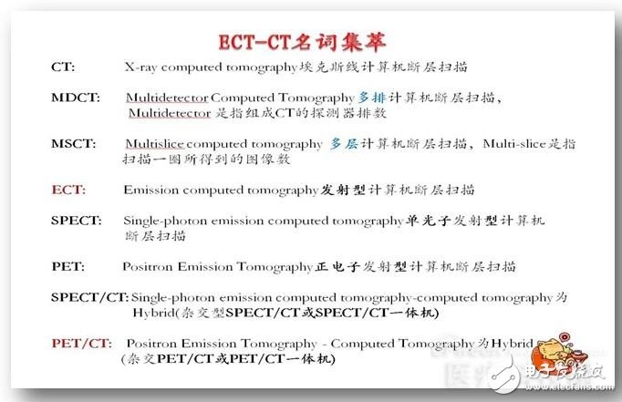
Speaking of PET and PET-CT, ECT is naturally a topic that can't be avoided. Strictly speaking, both PET and SPECT belong to the ECT family members. However, due to historical habits, ECT is generally referred to as SPECT in China, and PET-CT is treated separately by everyone because of its "expensive" and "new". The following nouns list the differences and connections between them.
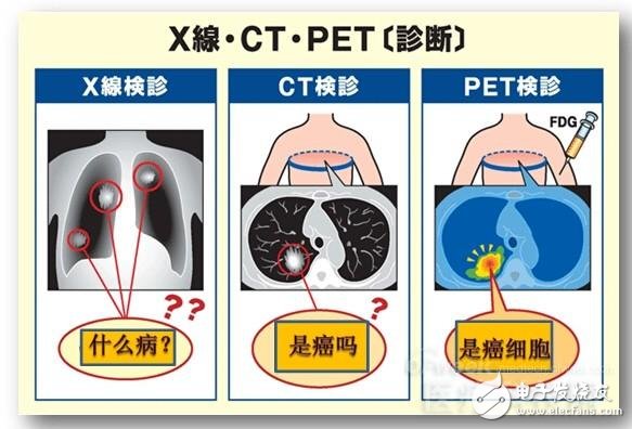
First of all, the working principle and composition of the "artifact" after PET image inspection, that is, how it works.
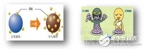
PET is a imaging device that reflects the genetic, molecular, metabolic, and functional states of a lesion. It uses positron-emitting radionuclides to label human compounds or metabolites, such as glucose, proteins, nucleic acids, fatty acids, receptor ligands, and human metabolites such as water as imaging agents. The most widely used clinical application is 18F- FDG, after injection into the subject, the positron nuclides accompany the labeled substance to participate in the cellular metabolic activities of human tissues and organs in the physiological and pathological process, and then re-aggregate and distribute, and then use PET to explore the external artifacts. In order to obtain information on the molecular level of metabolic activity of the human body, to provide clinical information on the biological metabolism of the disease. This process is like sending a spy inside the "enemy" (disease), and its internal situation is sent by a radio device carried by the spy, using the radio. Professional acceptance of equipment, the internal situation of the enemy can be clearly and timely grasped.
The whole set of PET equipment hardware consists of PET gantry, detector ring, electronic circuit and computer system, and image workstation. The software section includes image reconstruction and image processing systems.
The positron-emitting radionuclides injected into the human body undergo β+ decay to produce positrons, and the positrons and electrons in the tissue are quenched, producing two pairs of photons with 511 keV but flying in opposite directions.
PET uses its enclosed surround detector array to make these back-to-back photons conformal measurements (ie, electron collimation), consistent with measurements to form a projection line map (SINO map).
The original conforming data is spatially classified and recombined to form a SINO map and sent to subsequent PET image reconstruction software for processing. The image reconstruction is performed on a computer, and the projection SINO map data is used to reconstruct a radionuclide distribution tomogram of the tissue or organ to be tested.
The reconstructed tomograms are combined to obtain a three-dimensional image.
The functions of the computer and workstation and the corresponding software packages are to complete data acquisition, system monitoring, image reconstruction and image processing, and to achieve various clinical operations and diagnostic requirements.
After talking about PET, then come back to the spoiler's work.
In 1967, Hounsfield conceived a CT scanner that was patented in 1968. By 1972, he developed a CT that could complete a tomographic image in less than five minutes. In 1973, an X-tomography scanner (XCT) was made.
CT is an X-ray computed tomography (X-Ray computed tomography), which is a familiar X-ray tomography technique. X-rays are generated by an X-ray generator, transmitted to the human body, and detected by the detector to penetrate the human body. The amount of X-rays, after data classification, computer processing and reconstruction, obtain human tomographic images, ie human body profiles, which can observe the morphological characteristics (anatomical structure, shape, size, density) of specific parts (or precise parts) of the human body in detail. Shows the specific characteristics of the lesion. Just like cutting a piece of watermelon, you can visually see how the color of the watermelon is cut, whether it is seeded or seedless. Where is the watermelon seed growing, where is the rotten piece, how big, how it looks, and It is clear that the boundary is not bad.
Now we will introduce PET/CT.
PET/CT integrated machine, namely positron emission tomography and X-ray computed tomography imaging system, integrates PET and CT organically, and combines them into one. The patient can obtain CT anatomical images and PET functional metabolic images at the same time. The two images are organically combined and complement each other to produce the perfect effect of 1+1"2. The biological metabolic information of the lesion and the precise anatomical positioning and structure are presented to the doctor at the same time, so that the doctor can judge the disease earlier and faster. More complete and more accurate.
Although the nuclides on the tracer used in PET have certain radioactivity, the doses used are extremely low, and they are all short-lived, and the half-life is very short. For example, commonly used 18F, 11C, etc., through their own attenuation and metabolism, soon Cleared from the body, so the radiation hazard is very low.
Usually, patients often encounter patients and ask: "How much does it cost to do a full-body CT examination?" It really makes people feel dumbfounded. CT scans are performed by sub-site and sub-scanning (flat or enhanced or reconstructed). No one will do a head-to-toe whole body scan. The artifact of the whole body scan does exist, that is, PET-CT, from head to toe, the body's various organ functions, metabolism and other physiological and pathological features, 20 minutes panoramic view. Obviously, PET, which focuses on the genetic, molecular, metabolic and functional state of the lesion, is a new milestone in the development of life science and medical imaging technology.
Sometimes, you think that your body is good like this, the face is rosy and shiny, in fact, it is already like this. It has already begun to have early reaction of the lesion. The advantage of PET is that when the apple looks good, you can see it. The bug inside is wriggling.
If PET is like a human body to check the navigation mark in the sea, then CT is the navigation map for determining the navigation mark. PET and CT combine to achieve accurate and fast targets. PET/CT has an advantage in guiding the diagnosis and treatment of tumors, heart and brain diseases.
The advantages of the combination of PET and CT are complementary, mutual cooperation and mutual control. PET combined with CT improves the accuracy of lesion localization; CT combined with PET improves the qualitative diagnosis of lesions; meanwhile, CT images for attenuation correction can greatly shorten the examination time.
The combination of PET and CT, since retaining the classic anatomical image and introducing advanced molecular imaging, PET/CT has an advantage in guiding the diagnosis and treatment of tumors, heart and brain diseases.
The PET/CT core is fusion. Image fusion refers to the image transformation of the same or different imaging methods to match their spatial and spatial coordinates. The image fusion processing system utilizes the characteristics of the respective imaging modes for the two images. Spatial registration and combination are performed to register image data into a single image. PET/CT has the same positioning coordinate system with the same machine. When the patient scans without changing the position, PET/CT can be collected in the same machine, which avoids the error caused by patient displacement.
PET/CT reconstructed images can simultaneously display transverse, coronal, sagittal, and any beveled layers, and can be used to change the location and thickness of the reconstruction to provide more information to the clinician.
In the PET/CT system, the patient's planar image and tomographic image are acquired by CT scan and transmitted to the PET subsystem to provide bed planning for subsequent PET scans, providing the basic "positioning image" and "attenuation image" required for PET attenuation correction. The PET image is subjected to attenuation correction to complete the fusion of the PET image and the CT image. The merged PET/CT image data is transmitted to the image fusion/clinical analysis workstation. The workstation has a variety of clinical processing protocols, which can facilitate the processing, diagnosis and quantitative analysis of patient image data, and has DICOM3 output function. The software interface of this product is all English interface, but there is a Chinese report system, which can print reports and film.
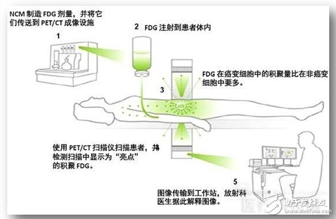
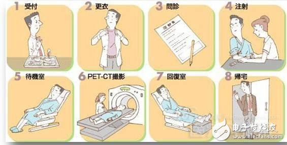
The main inspection process for PET-CT is shown in the figure below, and the specific details are implemented according to the specific requirements of the inspection structure.
Features of Nomex Braided Sleeving
long-term working temperature: -40 ° C -360 ° C
This Braided Expandable Sleeving is lightweight, flexible and non-flammable, it will perform without deterioration in the most diverse and demanding environments.
Nomex braided sleeving is oil and water repellent and also offers excellent resistance to gamma and x-rays.
Nomex is designed for the protection of wires and cable bundles against flame, high temperatures and mechanical abrasion. It is used on a variety of applications in the military, railway, marine, aeronautical and electrical equipment industries.
1: 3 expansion ratio
• Thin Wall
• Lightweight
• Self-extinguishing
• Excellent abrasion protection
With a tensile strength of 90,000 psi and can withstand temperatures up to 700°F, the Nomex Braided Sleeving is known for its high flame resistance. Because of the smooth and soft material, Nomex is used to make fire resistant protective gear for individuals that work in extreme heat conditions.
Nomex Braided Sleeving
Nomex Cable Sleeve,Armid Nomex Wire Braided Sleeve ,High Temperature Wire Sleeve,Heat Braided Sleeve For Wire
Shenzhen Huiyunhai Tech.Co.,Ltd , https://www.hyhbraidedsleeve.com